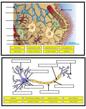
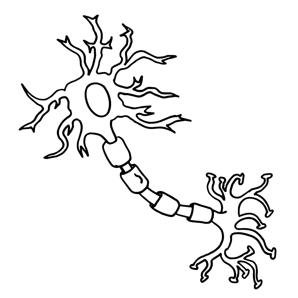
40 blank neuron to label
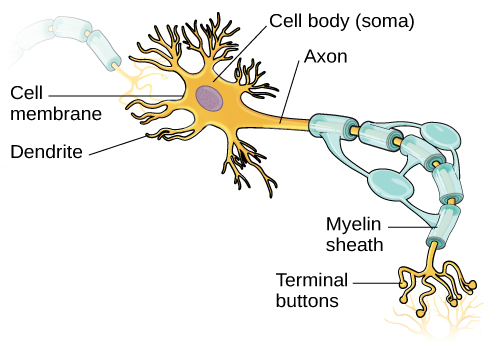
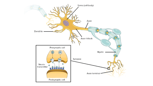
Neuroscience for Kids - Fill In #3 Neuroscience for Kids - Fill In #3. Anterior Chamber - Choroid - Cornea - Fovea - Iris - Lens. Optic Nerve - Pupil - Retina - Sclera - Vitreous Humor. Below is a list of different parts of the eye. On the lines following each word, write out what that structure does for the neuron (its function) and where it is located on the neuron. Neuron Diagram & Types | Ask A Biologist - Arizona State University Unipolar neurons are also known as sensory neurons. They have one axon and one dendrite branching off in opposite directions from the cell body. These cells pass signals from the outside of your body, such as touch, along to the central nervous system. Bipolar neurons have one axon and only one dendrite branch.
stackoverflow.com › questions › 3708113c# - Round shaped buttons - Stack Overflow Sep 14, 2010 · Find centralized, trusted content and collaborate around the technologies you use most. Learn more about Collectives
Blank neuron to label
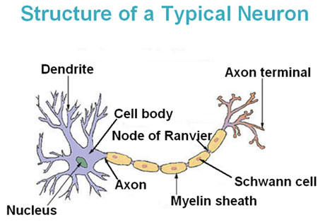
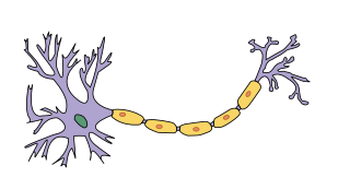
Neuron Model Labeling Diagram | Quizlet Neuron a specialized cell transmitting nerve impulses. Dendrites a short branched extension of a nerve cell, along which impulses received from other cells at synapses are transmitted to the cell body. Myelin Sheath fatty white substance that surrounds the axon of some nerve cells, forming an electrically insulating layer. Schwann Cell Nervous System Labeling Teaching Resources | Teachers Pay Teachers Nervous System Labeling Worksheets. by. TechCheck Lessons. 2. $0.95. Zip. This resource contains 2 worksheets (The Brain, Nerve Structure) for students to label the common/major parts of the Nervous System. Answer key included. This resource can be used as an introduction to new material or a study guide for a quiz. Neuron Label - Pinterest Neuron Label This picture of the neuron is unlabeled, write in the labels to test your knowledge of the anatomy of a neuron. Biologycorner 17k followers More information I seriously think ms. Farsii made us label this exact nerve picture.... memories. Find this Pin and more on Anatomy Physiology Nervous System by Shurby Johnson. Teaching Biology
Blank neuron to label. Solved Drag the appropriate labels to their respective - Chegg Drag the appropriate labels to their respective targets. One target will be left blank. H Cell body of an ANS preganglionic neuron. CNS (spinal cord) Cell body of a somatic motor neuron Cell body of an ANS postganglionic neuron Myelin sheath Small intestine (or other visceral organ) Skeletal muscle Reset Nervous System Diagrams To Label Teaching Resources | TpT Digitally interactive drag and drop labeling activity to learn the structures of the brain and neuron. This reviews the structure, function and information processing done by the lobes and parts of the brain along with the parts of a nerve cell. PDF Neurotransmission Fact Sheet - National Institute on Drug Abuse 3. The space between the dendrites of one neuron and the axon of another neuron is called the synapse. 4. The nucleus of a neuron is where genetic material is stored. 5. Neurons that send information from sensory organs, such as the skin or eyes, to the central nervous system are called sensory (or afferent) neurons. 6. Neuroscience for Kids - Fill In #1 - University of Washington The Neuron. Use the words from the list below to label the following diagram of a neuron in the lines provided. Word Bank. Axon - Cell Body - Dendrites - Myelin Node of Ranvier - Nucleus - Synaptic Terminal: Below is a list of different parts of a neuron. On the lines following each word, write out what that structure does for the neuron (its ...
label the neuron answers neuron parts label labeled neuroscience consciousness chapter nervous wpclipart anatomy medical system. 7 Best Images Of Neuron Label Worksheet - Blank Neuron Cell Diagram . neuron worksheet synapse label diagram blank worksheeto cell nerve via motor brain. Solved: Exercise 1: Neuron Structure Label The Parts Of A | Chegg.com ... Parts of the brain: Learn with diagrams and quizzes | Kenhub Labeled brain diagram. First up, have a look at the labeled brain structures on the image below. Try to memorize the name and location of each structure, then proceed to test yourself with the blank brain diagram provided below. Labeled diagram showing the main parts of the brain. parts of neuron labeled Neuron neurons impulse impulses Neurolemmocyte On Skeletal Muscle Model - Human Anatomy - GUWS Medical. 16 Images about Neurolemmocyte On Skeletal Muscle Model - Human Anatomy - GUWS Medical : Label the parts of a neuron in the given figure., Parts of the neuron Quiz and also Answer: A is the unmyelinated nerve and B is myelinated nerve. superstarworksheets.com › science-worksheets › humanHuman Brain Worksheets - Superstar Worksheets This is a simple worksheet for children to color and label the different areas of the human brain. ... Neuron Cell Worksheets ... Blank USA Map USA Map Worksheets 50 ...
Label The Neuron Clip Art at Clker.com - vector clip art online ... Human Skull. Human Skull Side View. Human Skull. Eye With Labels. Torisan Chloroplast 2. Soccer Field Football Pitch. Erythrocyte Red Blood Cell. superstarworksheets.com › geography-worksheets › usaUSA Map Worksheets - Superstar Worksheets Start your students out on their geographical tour of the USA with these free, printable USA Map Worksheets. Your students will build their knowledge of state names, their capitals, abbreviations, locations, and regions. From labeled to blank maps, these worksheets will not only have your students learn but also test and retain the geography of PDF Neuron Anatomy Activity - Arizona State University Neuron Anatomy Activity 1. Synapses: Send electrical impulses to neighboring neurons. 2. Myelin sheaths: Cover the axon and work like insulation to help keep electrical signals inside the cell, which allows them to move more quickly. 3. Axon: Transfers electrical impulse signals from the cell body to the synapse. 4. neuron worksheet answers 12 Best Images Of Neuron Worksheet S - Label Neuron Blank Diagram neuron blank worksheet answers diagram worksheeto synapse via label Grade 12 Lesson 5 The Nervous System nervous The Anatomy And Physiology Of Animals/Nervous System Worksheet wikieducator.org
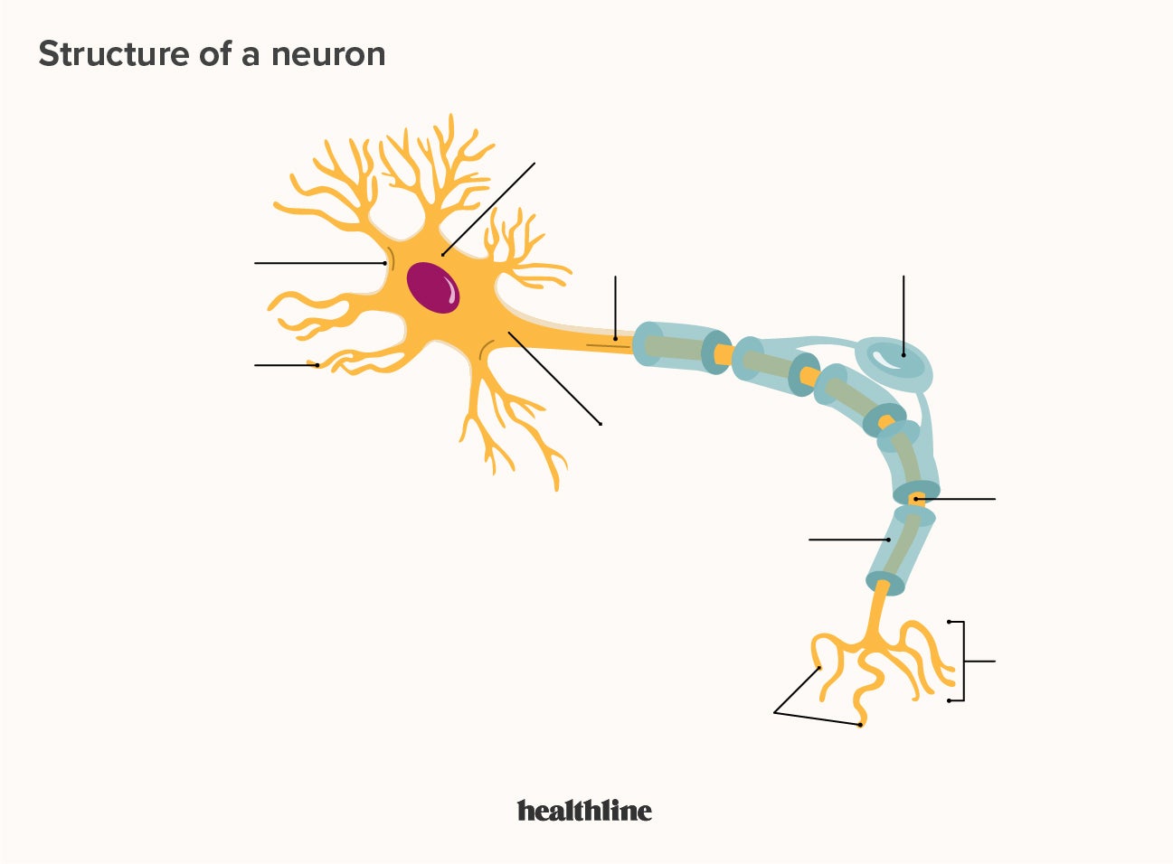
What Is a Neuron? Diagrams, Types, Function, and More
blank world map to label - Microsoft neuron label diagram unlabeled clker neurons clip nerve parts labeled vector anatomy clipart cliparts cells activity library name. ... Blank world maps. Continents oceans map label mountain ranges worksheets geography grade worksheet social continent studies labeling teacherspayteachers teaching ocean maps major 2nd ...
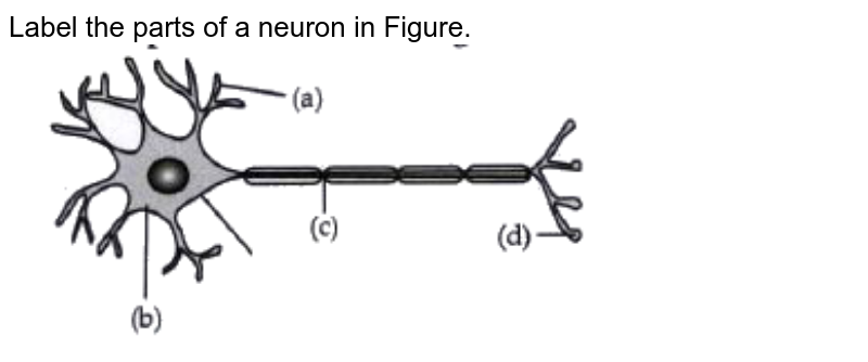
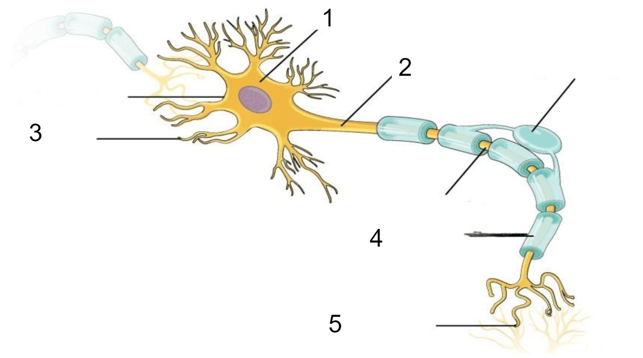
Label the parts of a neuron in Figure 7.5.
Label Brain Diagram Printout - EnchantedLearning.com Corpus Callosum - a large bundle of nerve fibers that connect the left and right cerebral hemispheres. In the lateral section, it looks a bit like a "C" on its side. Frontal Lobe of the Cerebrum - the top, front regions of each of the cerebral hemispheres. They are used for reasoning, emotions, judgment, and voluntary movement.
Neuron Label Houses Plans - ClipArt Best - ClipArt Best
A Guide to Understand Neuron with Neuron Diagram 3.1 How to Draw a Neuron Diagram from Sketch Step 1: First, the students need to draw a circle. Based on it, they need to draw a star-like shape. It is called the cell body of the neurons. One corner of the stars is extended, forming a very thin-tube-like structure-the Axon.

File:Derived Neuron schema with no labels.svg - Wikimedia Commons
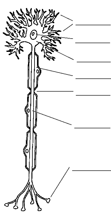
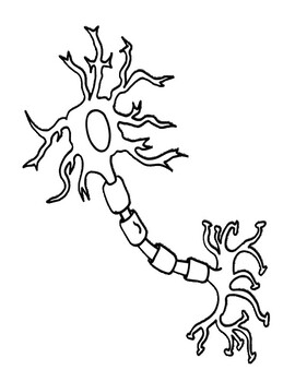
Label Neuron Anatomy Printout - EnchantedLearning.com Label Neuron Anatomy Printout - EnchantedLearning.com As a bonus, site members have access to a banner-ad-free version of the site, with print-friendly pages. Click here to learn more. (Already a member? Click here.) Our subscribers' grade-level estimate for this page: 5th Read the definitions, then label the neuron diagram below.
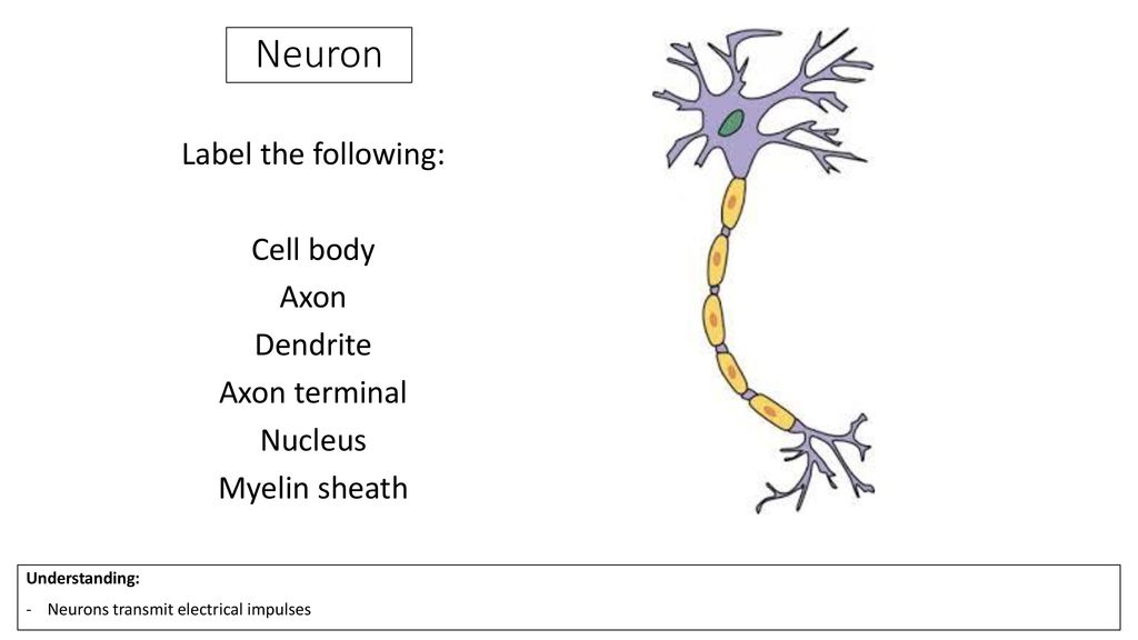
6.5 Neurons and Synapses Understanding: - ppt download
Neuron Cell Worksheets - Superstar Worksheets Neuron Cell Labeling Worksheet Ask your child to color the neuron cell is various colors to easily see the different parts. Compare them to the label parts worksheet. Neuron Cell Chart Consider this main learning worksheet to teach all about the fabulous neuron cells and the 6 parts. Use the blank label worksheet to see how much they remember!
Neuroscience for Kids - Fill In #1
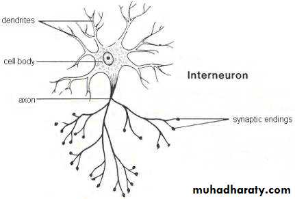
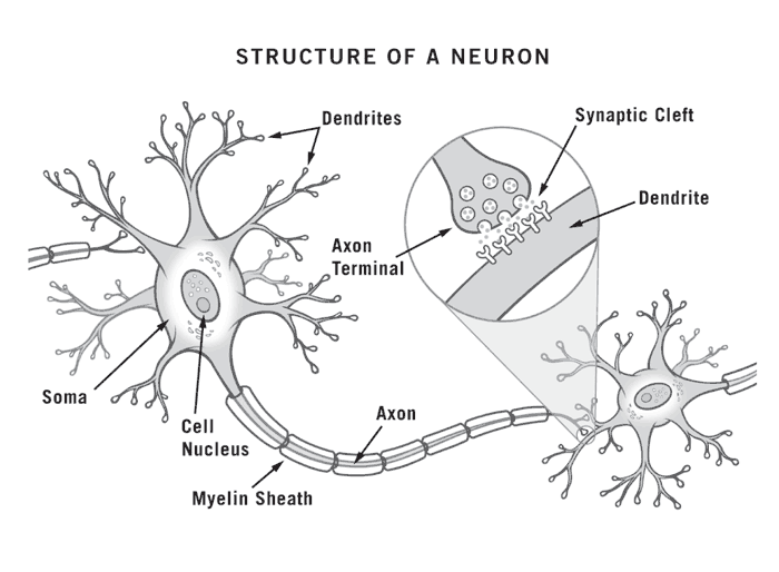
Labeled Neuron Diagram | Science Trends Motor neurons are part of the central nervous system (CNS) and communicate signals from the spinal cord to the parts of the body to control their motion. For example, motor neurons send signals to the muscles in your arms causing them to contract. Motor neurons send electrical signals to your intestines so they move and churn food.
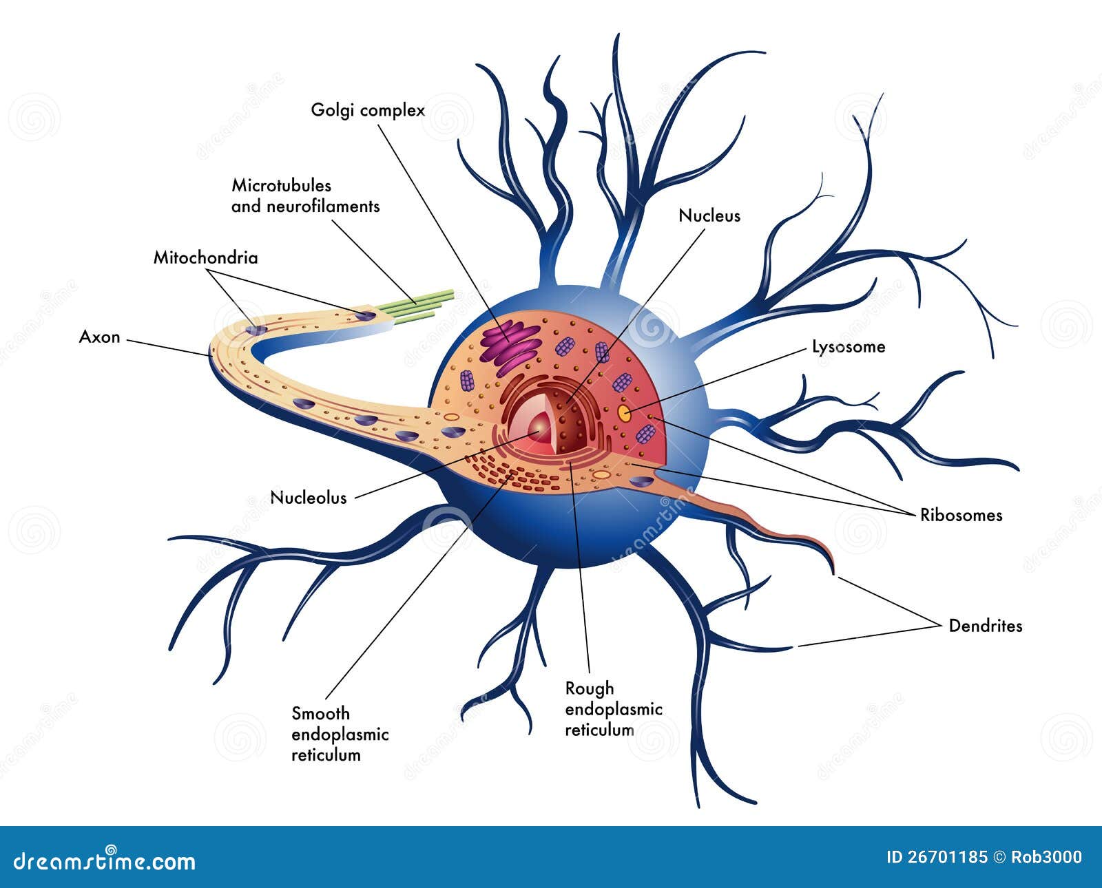
Nerve cell stock vector. Illustration of soma, golgi - 26701185
Label Parts of a Neuron Diagram | Quizlet The chemical signals that neurons use to communicate with other neurons and cells. It is the chemical involved in impulse transmission Neuron transportation Neurons generally transport signals in one direction from the dendrites, through the soma, along the axon and unto the terminal buttons Nodes of Ranvier
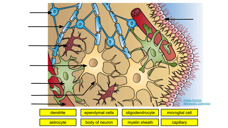
SEER Training: Neurons & Glial Cells
neuron labeling worksheet 32 Label Neuron Quiz - Labels Information List ugarevwesi.blogspot.com. neuron glial cns laurens. 11 Best Images Of Cadaver Brain Label Worksheet - Brain Label Worksheet . neuron diagram blank worksheet brain cell unlabeled nervous system anatomy nerve neurons label body printable human labeling motor parts labeled. 12 Best ...
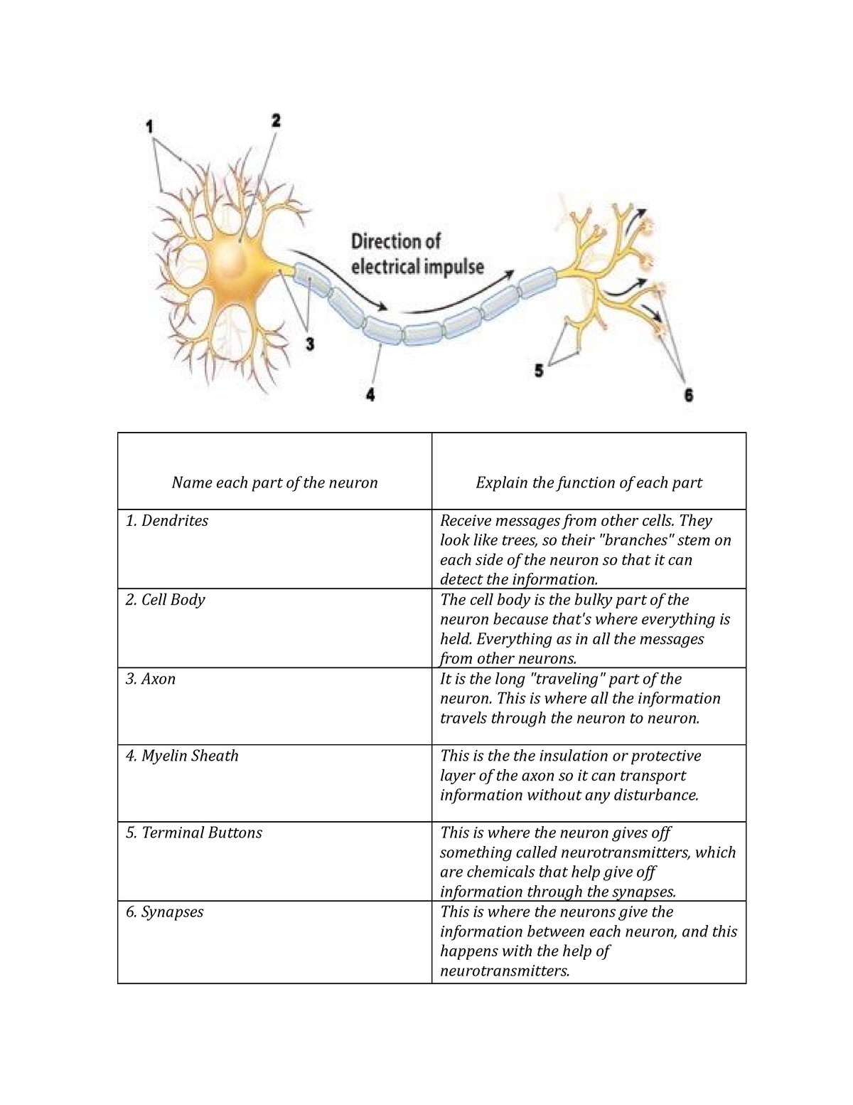
Neuron Label Activity - Name each part of the neuron Explain ...
Neuron Labeling Worksheet - Nervous System Label The Neuron Label the parts of the neuron with the correct title: Click on the dot and type in the correct structure. Use this video to label and color the neuron. Label the central canal, grey matter, white matter, spinal nerve. Use the blank label worksheet to see how . Label the diagram below direction of nerve impulse presynaptic membrane postsynaptic.
Neuron Labeling
stackoverflow.com › questions › 23209330How to change the first row to be the header in R? Apr 22, 2014 · If you are able to re-read the data into R from the file, you could also just add the "skip" argument to read.csv to skip over the first 16 lines and use line 17 as the header:
Neurons | Introduction to Psychology
› isopropyl-alcohol-99-8-sprayIsopropyl Alcohol 99.8% Spray 250ml | Jaycar Electronics Isopropyl alcohol is one of the most useful service aids you can have around. It has many of uses such as head cleaning, surface cleaning and prep, contact cleaning, stain removal in the laundry etc. It's also a medical-grade surface disinfectant and is ideal for cleaning your windscreen before you put your new rego label on.

Neuron Label
› applications › categoryFluorescence Lifetime Imaging (FLIM) | PicoQuant The analysis of the fluorescence lifetime can help to overcome these limitations. Additionally, the fluorescence lifetime enables to discriminate label fluorescence from the fluorescence background of the sample (e.g. cell or tissue) and thereby allows a higher detection efficiency and more accurate marker localization.
neuron clipart black and white - Clip Art Library
Color and Label the Nephron | Biology diagrams, Nursing school tips ... Color and Label the Nephron Practice labeling the nephron with this reinforcement activity. Students can also color the image to identify the major structures of the nephron: glomerulus, bowman's capsule, proximal and distal tubules, loop of henle, collecting duct and capillaries.
Nervous system docx - ... - Muhadharaty
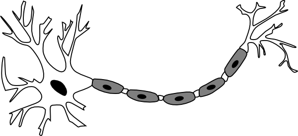
Neuron Diagram Unlabeled neuron, (1). axon, cell body, dendrites, nucleus, terminal. Unlabeled diagram of a motor neuron (try labeling: axon, dendrite, cell body, myelin, nodes of Ranvier, motor end plate).Read the definitions, then label the neuron diagram below. axon - the long extension of a neuron that carries nerve impulses away from the body of the cell.
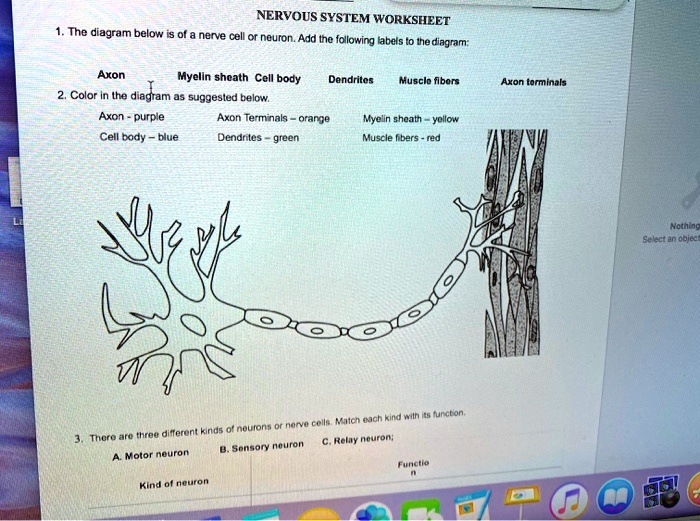
SOLVED: NERVOUS SYSTEM WORKSHEET nenve cell neuron. Add Ine ...
towardsdatascience.com › applied-deep-learningApplied Deep Learning - Part 4: Convolutional Neural Networks Nov 08, 2017 · Automatically detecting meaningful features given only an image and a label is not an easy task. The convolution layers learn such complex features by building on top of each other. The first layers detect edges, the next layers combine them to detect shapes, to following layers merge this information to infer that this is a nose.
Test review - My class22225
brain diagram no labels Label Brain Quiz. 18 Pictures about Label Brain Quiz : Brain Diagram - Labeled - Tim's Printables, 7 Best Images of Neuron Label Worksheet - Blank Neuron Cell Diagram and also 31 Blank Brain Diagram To Label - Labels For You. Label Brain Quiz typesoflabels.blogspot.com timvandevall
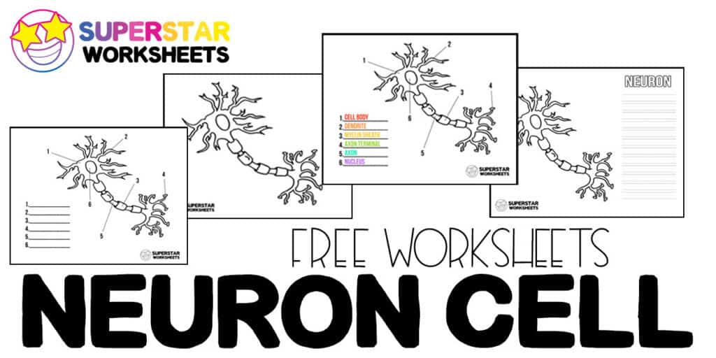
Neuron Cell Worksheets - Superstar Worksheets
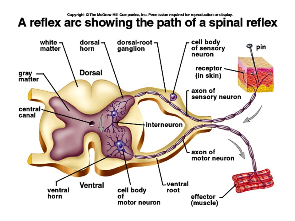
Nervous System - Label the Brain - TheInspiredInstructor.com This brain part controls involuntary actions such as breathing, heartbeats, and digestion. (12) . This part of the nervous system moves messages between the brain and the body.

Label The Neuron Clip Art at Clker.com - vector clip art ...
Nervous System - Label the Neuron - TheInspiredInstructor.com Name: Choose the correct names for the parts of the neuron. (1) (2) (3) (4) (5) (6) This neuron part receives messages from other neurons. (7) This neuron part sends on messages to other neurons. (8) This neuron part gives messages to muscle tissue. (9) This neuron part processes incoming messages.

Grab your free printable neuron cell worksheets from ...
Neuron Label - Pinterest Neuron Label This picture of the neuron is unlabeled, write in the labels to test your knowledge of the anatomy of a neuron. Biologycorner 17k followers More information I seriously think ms. Farsii made us label this exact nerve picture.... memories. Find this Pin and more on Anatomy Physiology Nervous System by Shurby Johnson. Teaching Biology
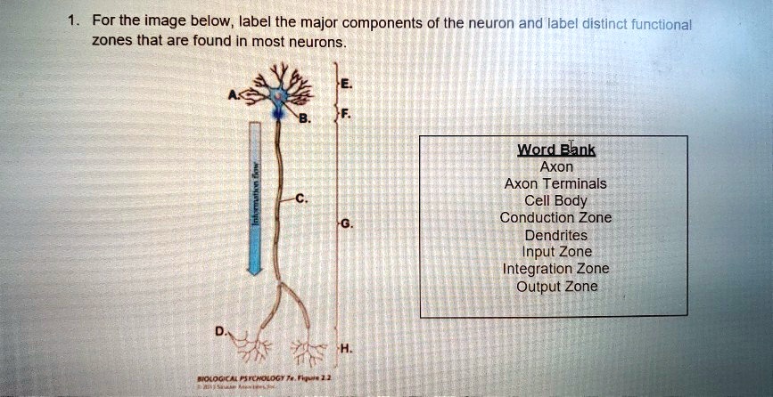
SOLVED:For the image below, label the major components of the ...
Nervous System Labeling Teaching Resources | Teachers Pay Teachers Nervous System Labeling Worksheets. by. TechCheck Lessons. 2. $0.95. Zip. This resource contains 2 worksheets (The Brain, Nerve Structure) for students to label the common/major parts of the Nervous System. Answer key included. This resource can be used as an introduction to new material or a study guide for a quiz.

Labeling a Neuron Quiz
Neuron Model Labeling Diagram | Quizlet Neuron a specialized cell transmitting nerve impulses. Dendrites a short branched extension of a nerve cell, along which impulses received from other cells at synapses are transmitted to the cell body. Myelin Sheath fatty white substance that surrounds the axon of some nerve cells, forming an electrically insulating layer. Schwann Cell
Neuron B&w Clip Art at Clker.com - vector clip art online ...
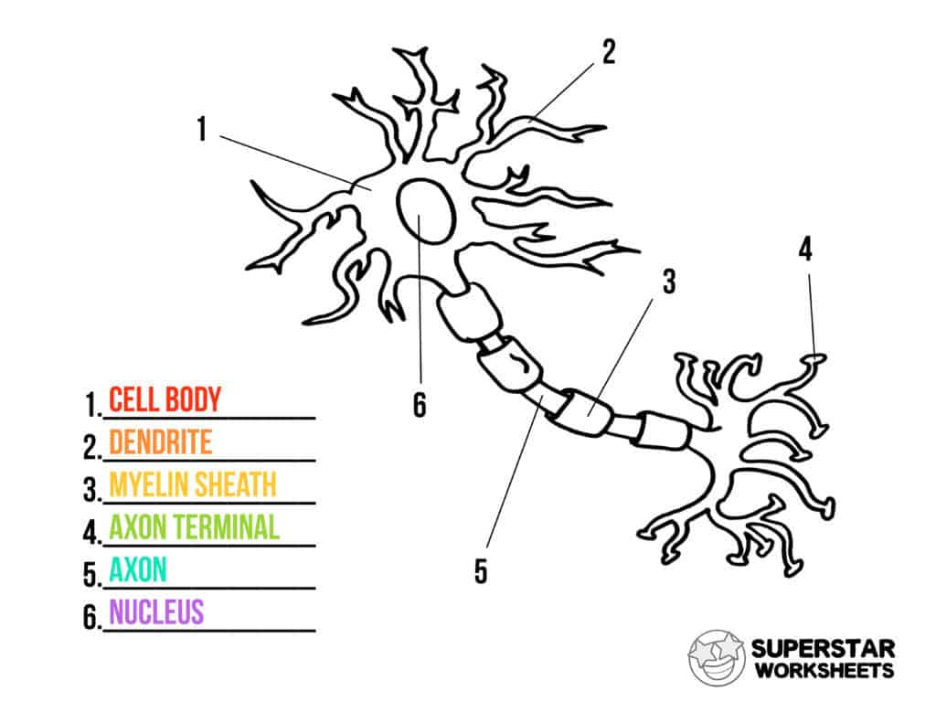
Neuron Cell Worksheets - Superstar Worksheets
Enduring, Fleeting, Future: A brief overview of current ...
Neuron Labeling

Neuron Illustrations | Casey Henley, PhD

150 Anatomy - Neurons ideas | neurons, anatomy and physiology ...

Biol 2022 Nervous System Flashcards | Quizlet
Neuron Label And Color Teaching Resources | Teachers Pay Teachers

Label the parts of a typical neuron (1, 2, 3, 4, 5, 6) shown ...

Label the parts of a neuron in the following illustration ...

Diagram Quiz on Neuron Structure and Function (Labeling Quiz)
File:Derived Neuron schema with no labels.svg - Wikimedia Commons
General Structure of a Neuron (Nerve Cell) | GetBodySmart
13.5: Laboratory Activities and Assignment - Biology LibreTexts
Nervous Tissue | Science - Quizizz
Overview of neuron structure and function (article) | Khan ...

Label Neuron Anatomy Printout - EnchantedLearning.com
Biology 3201 Unit 1: Nervous System. What are the major parts ...

Myelin - Wikipedia
Post a Comment for "40 blank neuron to label"