39 labeling a compound microscope
Compound Light Microscopes | Products | Leica Microsystems Aug 08, 2018 · A compound microscope uses optics to produce a magnified image of a sample so that with details of it can be observed that are undetectable with the naked eye. The most basic optics of a compound microscope has at least 2 lenses: i) an objective placed nearby the sample which creates a magnified, real image of it and ii) eyepieces or oculars ... How to draw compound of Microscope easily - step by step I will show you " How to draw compound of microscope easily - step by step "Please watch carefully and try this okay.Thanks for watching.....#microscopedrawi...
Microscope Cameras | Products | Leica Microsystems Jul 23, 2018 · Microscope Cameras for Industry Optimum visualization and ease-of-use Industrial applications require microscope cameras which: i) are capable of high frame rates for fast live images to accommodate a rapid workflow; ii) can provide excellent image quality for precise analysis; and iii) are easy to use even for inexperienced users.

Labeling a compound microscope
Labeling the Parts of the Microscope | Microscope World Resources Labeling the Parts of the Microscope. This activity has been designed for use in homes and schools. Each microscope layout (both blank and the version with answers) are available as PDF downloads. You can view a more in-depth review of each part of the microscope here. Compound Microscope - Diagram (Parts labelled), Principle and Uses As the name suggests, a compound microscope uses a combination of lenses coupled with an artificial light source to magnify an object at various zoom levels to study the object. A compound microscope: Is used to view samples that are not visible to the naked eye. Uses two types of lenses - Objective and ocular lenses. Label the microscope — Science Learning Hub All microscopes share features in common. In this interactive, you can label the different parts ...
Labeling a compound microscope. Microscope Labeling Practice Quiz - PurposeGames.com Practice labeling a compound microscope. Do you know all the parts? Remaining 0. Correct 0. Wrong 0. Press play! 0%. 0:00.0. Quit. Again. This game is part of a tournament. You need to be a group member to play the tournament. Join group, and play Just play. Your Scorecard. The scorecard of a champion. Score . 0 % Time Labeling a Compound Microscope Flashcards | Quizlet Start studying Labeling a Compound Microscope. Learn vocabulary, terms, and more with flashcards, games, and other study tools. Microscope Types (with labeled diagrams) and Functions Compound microscope labeled diagram. Compound microscope functions: It finds great application in areas of pathology, pedology, forensics etc; Its greater order of magnification allows for deeper study of microbial organisms to Detect the cause of diseases; Study the mineral composition in soils; Examine evidences collected in crime scenes by forensics. Labeling the Parts of the Microscope | Microscope activity, Science ... Jan 13, 2016 - Free worksheets for labeling parts of the microscope including a worksheet that is blank and one with answers. ... Description Worksheet identifying the parts of the compound light microscope. Answer key: 1. Body tube 2. Revolving nosepiece 3. Low power objective 4. Medium power objective 5. High power objective 6. Stage clips 7.
Compound Light Microscope: Everything You Need to Know A compound light microscope is a type of light microscope that uses a compound lens system, meaning, it operates through two sets of lenses to magnify the image of a specimen. It's an upright microscope that produces a two-dimensional image and has a higher magnification than a stereoscopic microscope. It also goes by a couple of other names ... Compound Microscope Parts, Functions, and Labeled Diagram The individual parts of a compound microscope can vary heavily depending on the configuration & applications that the scope is being used for. Common compound microscope parts include: Compound Microscope Definitions for Labels Eyepiece (ocular lens) with or without Pointer: The part that is looked through at the top of the compound microscope. Eyepieces typically have a magnification between 5x & 30x. Microscope Parts and Functions Before exploring microscope parts and functions, you should probably understand that the compound light microscope is more complicated than just a microscope with more than one lens. First, the purpose of a microscope is to magnify a small object or to magnify the fine details of a larger object in order to examine minute specimens that cannot ... Label a Compound Microscope Lesson Plans & Worksheets In this microscope worksheet, students complete 15 review questions of labeling, defining and short answer after finishing a compound microscope lab. Lab included. Get Free Access See Review
Microscope Labeling Game - PurposeGames.com About this Quiz. This is an online quiz called Microscope Labeling Game. There is a printable worksheet available for download here so you can take the quiz with pen and paper. This quiz has tags. Click on the tags below to find other quizzes on the same subject. Science. Microscopes for sale | eBay Compound microscopes: Compound microscopes are powerful microscopes that magnify items on glass slides. They have two or more convex lenses, and they can magnify objects by between 40 and 100 times. The images rendered by these microscopes are two-dimensional, and they are either monocular, binocular, or trinocular in design. Electron microscope - Wikipedia An electron microscope is a microscope that uses a beam of accelerated electrons as a source of illumination. As the wavelength of an electron can be up to 100,000 times shorter than that of visible light photons , electron microscopes have a higher resolving power than light microscopes and can reveal the structure of smaller objects. How to Use a Compound Microscope: 11 Steps (with Pictures) - wikiHow Focus the microscope. Looking through the eyepiece, arrange the illuminator and the diaphragm to reach the most comfortable level of light. Move the specimen slide so that the image is in the center of your view. [10] Arrange the illuminator until you've arrived at a comfortable level of light.
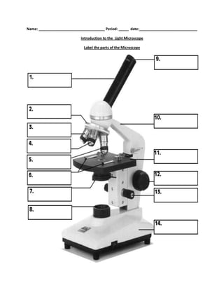
Label the microscope — Science Learning Hub
Labelled Diagram of Compound Microscope The below mentioned article provides a labelled diagram of compound microscope. Part # 1. The Stand: The stand is made up of a heavy foot which carries a curved inclinable limb or arm bearing the body tube. The foot is generally horse shoe-shaped structure (Fig. 2) which rests on table top or any other surface on which the microscope in kept.

Parts of Stereo Microscope (Dissecting microscope) – labeled ...
Parts of a Compound Microscope (And their Functions) - Scope Detective List of Microscope Parts and their Functions. 1. Ocular Tubes (Monocular, Binocular & Trinocular) The ocular tubes, are to tubes that lead from the head of the microscope out to your eyes. On the end of the ocular tubes are usually interchangeable eyepieces (commonly 10X and 20X) that increase magnification.
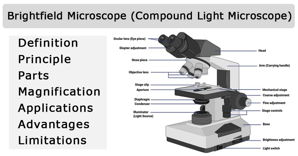
Brightfield Microscope (Compound Light Microscope ...
Parts of a microscope with functions and labeled diagram - Microbe Notes Head - This is also known as the body. It carries the optical parts in the upper part of the microscope. Base - It acts as microscopes support. It also carries microscopic illuminators. Arms - This is the part connecting the base and to the head and the eyepiece tube to the base of the microscope.

9 Compound Microscopes For Larger Magnification
Compound Microscope Labeled Diagram | Quizlet QUESTION. The total magnification of a specimen being viewed with a 10X ocular lens and a 40X objective lens is. 15 answers. QUESTION. a mosquito beats its wings up and down 600 times per second, which you hear as a very annoying 600 Hz sound. if the air outside is 20 C, how far would a sound wave travel between wing beats. 2 answers.

Microscope With Labels Clip Art at Clker.com - vector clip ...
Compound Microscope: Definition, Diagram, Parts, Uses, Working ... - BYJUS A compound microscope is defined as. A microscope with a high resolution and uses two sets of lenses providing a 2-dimensional image of the sample. The term compound refers to the usage of more than one lens in the microscope. Also, the compound microscope is one of the types of optical microscopes. The other type of optical microscope is a simple microscope.
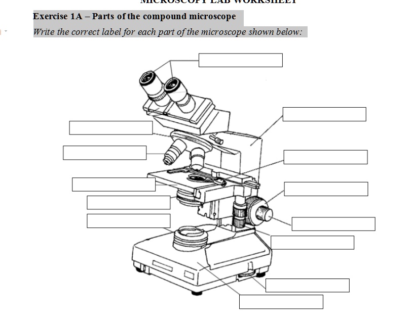
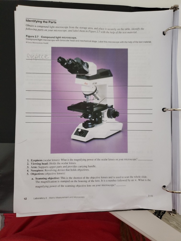
SOLVED: Exercise 1A _ Parts ofthe compound microscope Write ...
Looking at the Structure of Cells in the Microscope ... Many light-microscope techniques are available for observing cells. Cells that have been fixed and stained can be studied in a conventional light microscope, while antibodies coupled to fluorescent dyes can be used to locate specific molecules in cells in a fluorescence microscope. Living cells can be seen with phase-contrast, differential ...
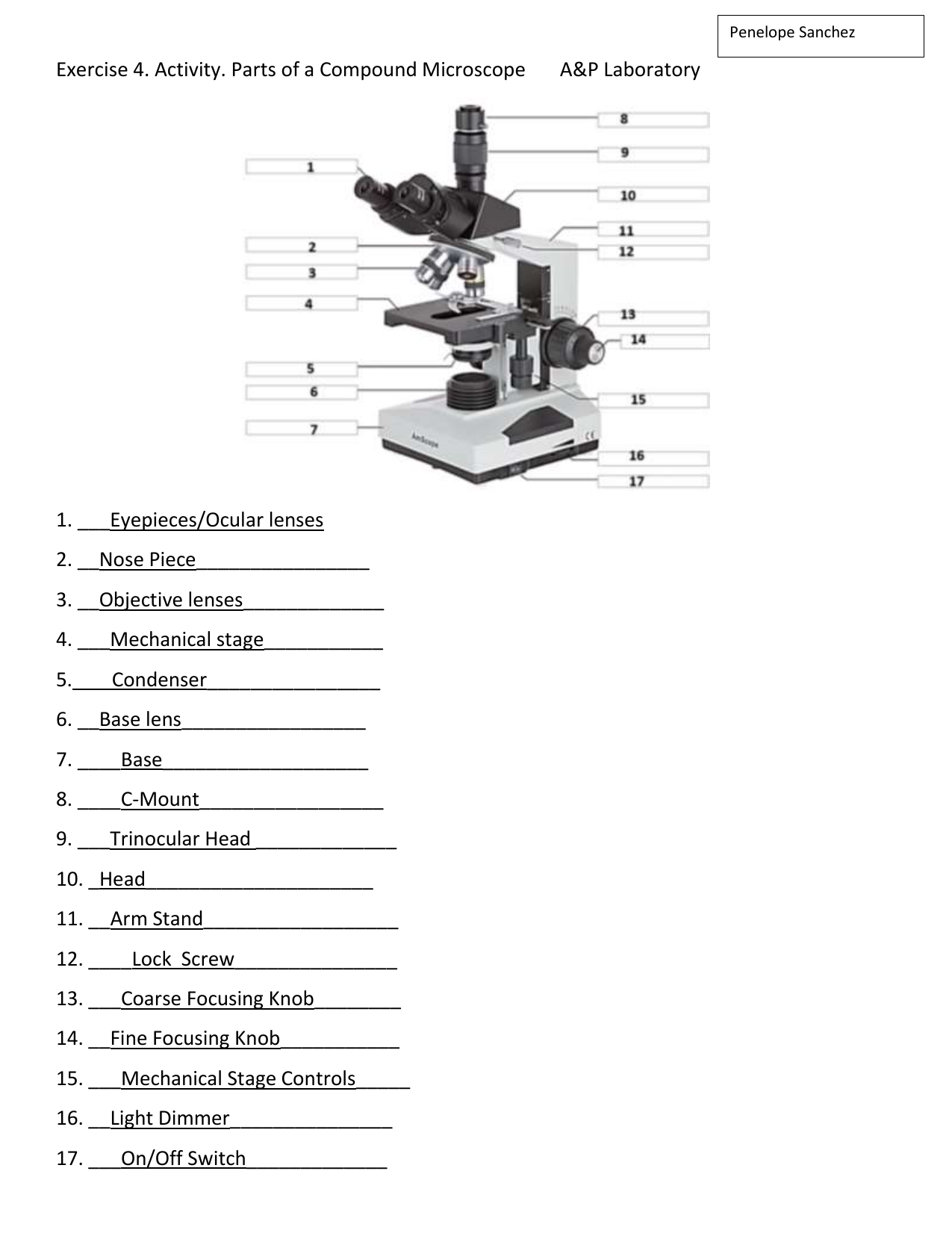
Exercise 4 Labeling Microscope Parts ACTIVITY d
Diagram of a Compound Microscope - Biology Discussion 1. It is noted first that which objective lens is in use on the microscope. 2. Stage micrometer is positioned in such a way that it is in the field of view. 3. The eyepiece is rotated so that the two scales, the eyepiece or ocular scale and the stage micrometer scale, are parallel. 4.

This is a common compound microscope. Label its parts from A ...
Microscope Labeling - The Biology Corner Microscope Labeling. Shannan Muskopf May 31, 2018. This simple worksheet pairs with a lesson on the light microscope, where beginning biology students learn the parts of the light microscope and the steps needed to focus a slide under high power. The labeling worksheet could be used as a quiz or as part of direct instruction where students ...
Biology label part of microscope
Drawing Of A Microscope And Label - Warehouse of Ideas The optical parts of the microscope are used to view, magnify, and produce an image from a specimen placed on a slide. Source: microspedia.blogspot.com. Learn how to draw microscope and label pictures using these outlines or print just for coloring. Controls the amount of light passing through the opening of the stage.

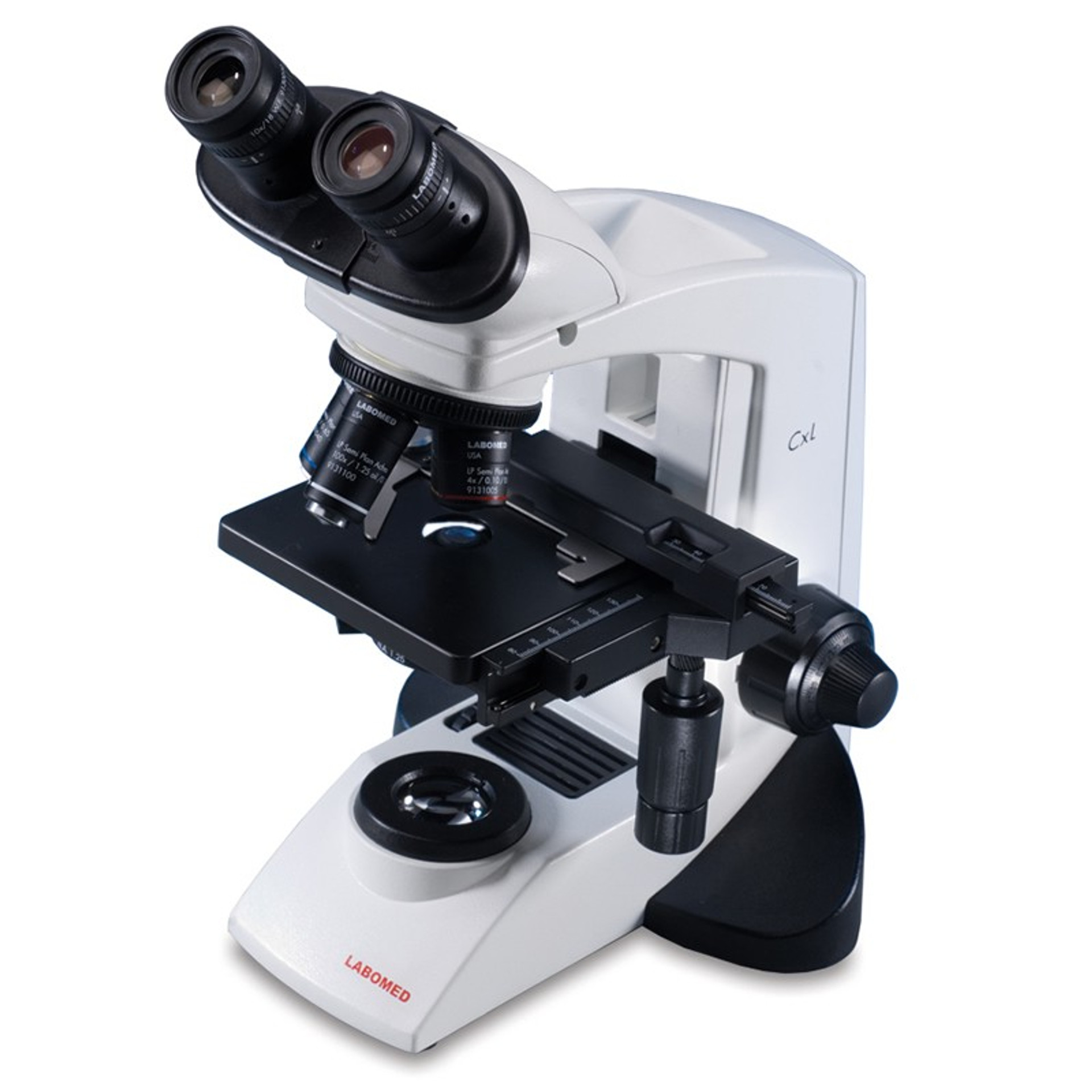
Amscope 40X-1000X Trinocular Biological Compound ...
ZEISS Axiolab 5 smart microscope for easy digital documentation Your smart microscope from ZEISS lets you assign microscope images with the correct scaling information to barcode-labeled samples. Just use your Axiolab 5 or Axioscope 5 microscope with Windows PC or iPad, connect a barcode reader to your Axiocam 208 color microscope camera and you’re good to go.

label the parts of the compound microscope - Brainly.ph
Parts of the Microscope with Labeling (also Free Printouts) 5. Knobs (fine and coarse) By adjusting the knob, you can adjust the focus of the microscope. The majority of the microscope models today have the knobs mounted on the same part of the device. Image 5: The circled parts of the microscope are the fine and coarse adjustment knobs. Picture Source: bp.blogspot.com.
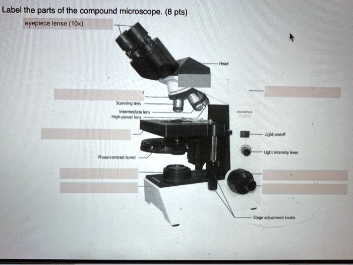
SOLVED:Label the parts of the compound microscope. (8 pts ...
Compound Microscope Parts - Labeled Diagram and their Functions There are two major optical lens parts of a microscope: Eyepiece (10x) and Objective lenses (4x, 10x, 40x, 100x). Total magnification power is calculated by multiplying the magnification of the eyepiece and objective lens. The illuminator provides a source of light. The light is focused by the condenser and passing through the specimen placed ...

COMPOUND MICROSCOPES & PARTS
Compound Microscope: Parts of Compound Microscope - BYJUS The parts of the compound microscope can be categorized into: Mechanical parts; Optical parts (A) Mechanical Parts of a Compound Microscope. 1. Foot or base. It is a U-shaped structure and supports the entire weight of the compound microscope. 2. Pillar. It is a vertical projection. This stands by resting on the base and supports the stage. 3. Arm

Biology - labeling a compound microscope Diagram | Quizlet
Labeling the Parts of the Microscope | Microscope World Resources Microscope World explains the parts of the microscope, including a printable worksheet for schools and home. Need Asssistance? 800-942-0528. Microscope Blog ... Labeling the Parts of the Microscope. This activity has been designed for use in homes and schools. Each microscope layout (both blank and the version with answers) are available as PDF ...

Simple Microscope - Diagram (Parts labelled), Principle ...
Compound Microscope - Types, Parts, Diagram, Functions and Uses Compound microscope - It has two convex lenses. It is called a compound microscope because it compounds the light as it passes through the lenses to magnify. The image of the object being viewed is enlarged because of the lens near the object. An eyepiece, an additional lens, is where real magnification takes place.

This is a common compound microscope Label its parts class 11 ...
label parts of a compound microscope - TeachersPayTeachers Students label the parts of a compound light microscope, match the part to it's function, watch a YouTube linked video on how to prepare a slide, and finally view a few simple specimens on their own.Materials needed:Compound Light MicroscopeMicroscope slides and cover platesMagazine/Newspaper letters to cut out and viewAnimal hair to view (I use dog or cat hair)Nylon fibers (such as from a rope)Wool fibers (such as from an old sock)Student device to take picturesThis file can be downloaded as a

Parts of a Microscope - SmartSchool Systems
Compound Microscope- Definition, Labeled Diagram, Principle, Parts, Uses The term "compound" in compound microscopes refers to the microscope having more than one lens. Devised with a system of combination of lenses, a compound microscope consists of two optical parts, namely the objective lens and the ocular lens.
Parts of a microscope with functions and labeled diagram
Label the microscope — Science Learning Hub All microscopes share features in common. In this interactive, you can label the different parts ...

Compound Microscope Parts – Labeled Diagram and their ...
Compound Microscope - Diagram (Parts labelled), Principle and Uses As the name suggests, a compound microscope uses a combination of lenses coupled with an artificial light source to magnify an object at various zoom levels to study the object. A compound microscope: Is used to view samples that are not visible to the naked eye. Uses two types of lenses - Objective and ocular lenses.

Parts of a Compound Microscope - Labeled (with diagrams ...
Labeling the Parts of the Microscope | Microscope World Resources Labeling the Parts of the Microscope. This activity has been designed for use in homes and schools. Each microscope layout (both blank and the version with answers) are available as PDF downloads. You can view a more in-depth review of each part of the microscope here.

Diagram of a Compound Microscope

Compound Microscope – Diagram (Parts labelled), Principle and ...

Compound Microscope Parts, Functions, and Labeled Diagram ...

Monday 10/19/15 AIM: how do the parts of the compound light ...

Compound Microscope Labeled Diagram | Quizlet

Compound Microscope: Parts of Compound Microscope

What is a Compound Microscope? | Microscope World Blog

Parts of a microscope with functions and labeled diagram

General Biology | Carlson Stock Art | General biology ...

LABELING THE COMPOUND LIGHT MICROSCOPE 2 Diagram | Quizlet

Labeled Microscope Diagram | Microscope parts, Science fair ...
parts of microscope drawing - Clip Art Library
Solved biology 1000 lab 2.7 compound light microscope ONLY ...

PARTS OF MICROSCOPE| LEARN TO LABEL COMPOUND MICROSCOPE| JUST ...
Compound Microscope Parts, Functions, and Labeled Diagram ...

Monday, 12 November 2018Monday, 12 November ppt download

Microscope labeling
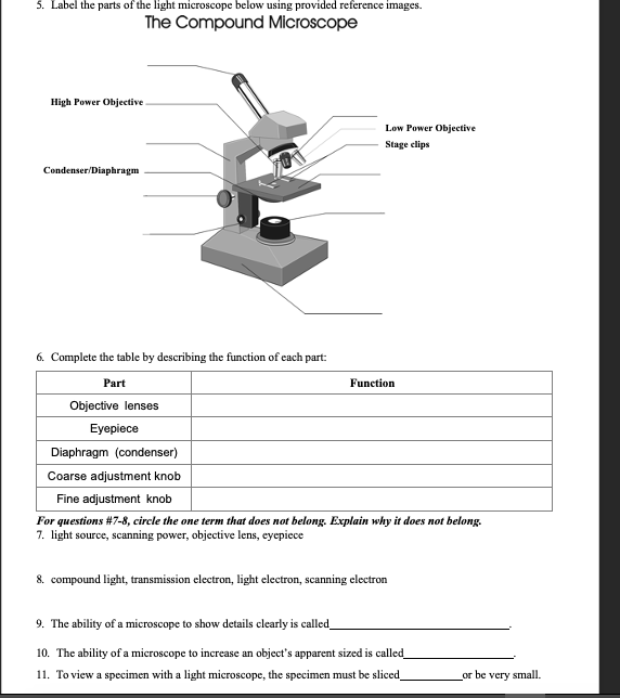
Solved 5. Label the parts of the light microscope below ...

National 131-LED-MS Compound Microscope 131-LED-MS B&H Photo
Post a Comment for "39 labeling a compound microscope"